Lithium-ion batteries have been widely applied in portable electronics due to their high energy densities. However, their potential applications in electric vehicles and grid energy storage call for higher energy density. It is a critical challenge to develop the next-generation electrochemical energy storage devices. Synchrotron X-ray imaging techniques are currently catching increasing attention due to their intrinsic advantages, including non-destructiveness, chemically responsiveness, elementally sensitivity, and high penetrability to enable operando investigation of a real battery. Based on the derived nano-tomography techniques, it can provide 3D morphological information including thousands of slice morphologies from the bulk to the surface. Combined with X-ray absorption spectroscopy, X-ray imaging can even present chemical and phase mapping information, including the oxidation state, local environment, etc., with sub-30 nm spatial resolution, which addresses the issues that we only obtain as averaged information in traditional X-ray absorption spectroscopy. Through an operando charging/discharging setup, X-ray imaging enables the study of the correlation between the morphology change and the chemical evolution (mapping) under different states of charge and cycling. In addition, X-ray imaging breaks up the size limit of nanoscale samples for the in-situ transmission electron microscope imaging, which enables a large, thick sample with a broad field of view, truly uncovering the behavior inside a real battery system. We will discuss a few major X-ray imaging technologies, including X-ray projection imaging, transmission X-ray microscopy, scanning transmission X-ray microscopy, tender and soft X-ray imaging, and coherent diffraction imaging. Researchers can choose from various X-ray imaging techniques with different working principles based on research goals and sample specifications. With the X-ray imaging techniques, we can obtain the morphology, phase, lattice and strain information of energy materials in both 2D and 3D in an intuitive way. In addition, with the high-penetration X-rays and the high-brilliance synchrotron sources, operando/in-situ experiments can be designed to track the qualitative and quantitative changes of the samples during operation. We expect this review can broaden readers' view on X-ray imaging techniques and inspire new ideas and possibilities in energy materials research.
Keywords:synchrotron radiation
;
X-ray
;
lithium-ion battery
;
the state of art characterization technology
AN Hanwen. Research progress of synchrotron radiation multimodal imaging technology in field of energy storage batteries[J]. Energy Storage Science and Technology, 2022, 11(3): 834-851
Fig. 2
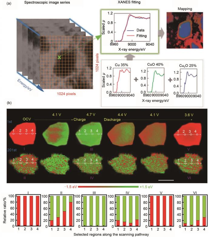
(a) working principles and data processing of TXM-XANES experiment on CuO anode[30]; (b) operando 2D spectroscopy imaging for characterization of single crystal LiNi0.6Co0.2Mn0.2O2 particles and corresponding charged state distribution statistics[22]
Fig. 3
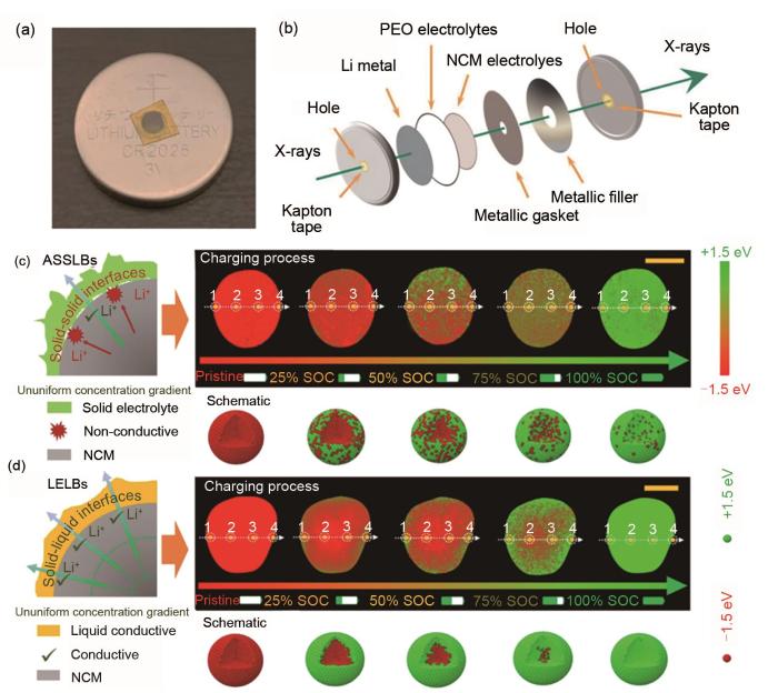
(a) solid polymer electrolyte cell; (b) schematic diagram of internal structure of solid-state battery model; (c) TXM-XANES mapping of single cathode particle as a function of charging time in ASSLBs; (d) corresponding schematic diagram to expound unique solid-state electrochemistry[33]
Fig. 4
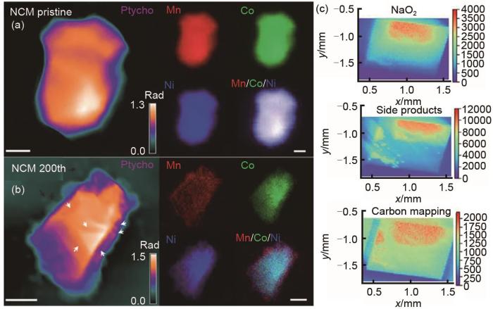
(a) X-rayfluorescence and ptychographic images of pristine; (b) 200th cycled NCM particles to show element distribution and morphology[22]; (c) X-rayfluorescence and ptychographic images of O and C[37]
Fig. 5
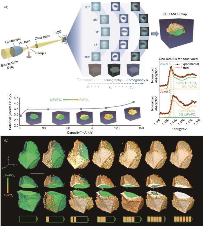
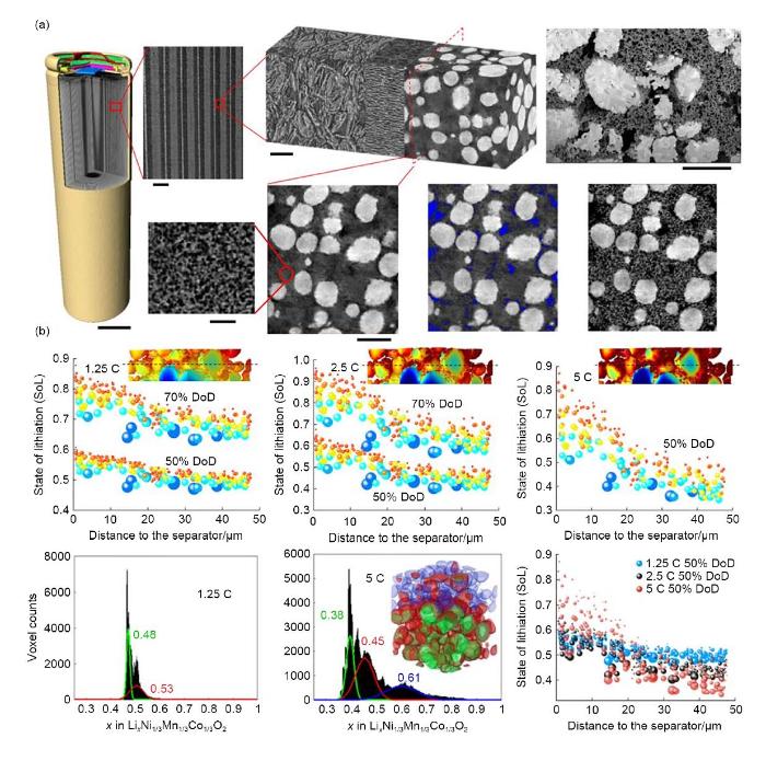
(a) 3D reconstructions of commercial 18650 NMC batteries[45]; (b) schematic illustration of experimental process for harvesting defective regions of interest from 18650-type Li-ion battery[46]; (c) 3D rendering of nanotomographic data of particle collected with four virtual slices[47]
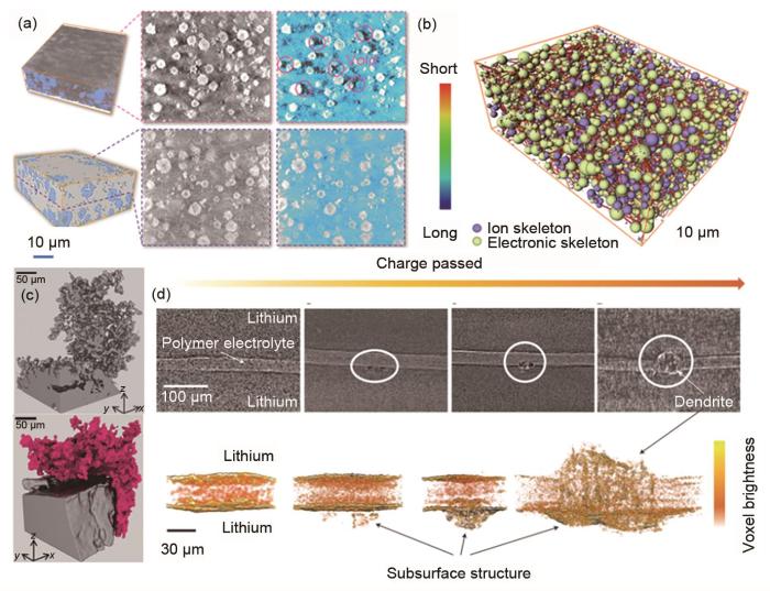
除了正极之外,同步辐射断层扫描技术也被应用于锂电池负极的研究。在锂电解质界面,人们普遍认为具有高弹性模量的固体电解质可以有效阻挡锂金属枝晶的生长[50]。然而,近期的研究表明锂离子仍能渗透固体电解质,最终使电池的循环稳定性和运行安全性恶化。枝晶引起的短路是开发高能量密度固态电池的最大瓶颈之一。虽然锂枝晶在固态电解质中生长的行为和机理还不完全清楚,但已证明紧密接触和均匀电流密度的固-固界面是均匀沉积锂的关键。在此基础上,本课题组[51]构建了三维Li x MnO2(LMO)电极骨架作为锂离子的快速输运通道,实现了Li的无枝晶溶解/沉积。利用同步加速器高分辨率X射线断层显微镜定量分析了电极的化学成分和孔隙率[图6(a)、(b)]。研究表明,LMO在电极内部的均匀分布可以形成均衡的离子输运网络,促进空间锂离子通量的均匀分布。在锂沉积后,发现空隙被Li金属完全填充,循环电极呈现密集的锂沉积。基于同步纳米CT数据的连通性网络模型也显示离子通量传输距离均匀且较短,进一步说明快速的锂离子传输是均匀镀锂的关键。与重元素相比,碳和锂等原子序数较低的元素对硬X射线的衰减较弱,导致它们与电解质填充的孔隙空间的对比度较低。随着相位对比技术的兴起,X射线显微断层扫描技术可以增强不同弱衰减材料(如碳和锂)之间的对比度。Shearing等[52]成功实现了锂枝晶的同轴相位对比成像,实现了衰减和折射效应与衰减相似材料之间的图像界面解耦[图6(c)]。相似地,图6(d)展示了X射线断层扫描技术对聚合物电解质/锂金属之间界面的研究[53]。
Fig. 6
(a) X-ray nanotomography and virtual slice of composite Li anode and cycled anode; (b) connectivity network of cycled composite Li electrode; intensity bar indicates Li+ transport distance[50]; (c) 3D rendering of Li microstructures under constant current (top), and higher density microstructures following plating and stripping (bottom); (d) Li dendrite growth with charge passed[52]
Fig. 7
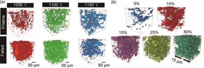
(a) morphological transformation of LLZO solid electrolytes between pristine and failed cells obtained by X-ray tomographic reconstructions; (b) reconstructed images for LLZO[20]
图8
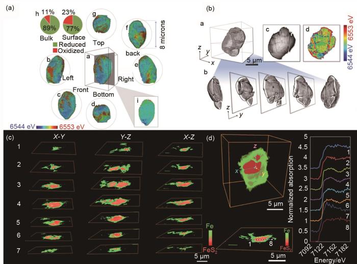
(a) Li x Ta0.3Mn0.4O2(LTMO)颗粒Mn价态的三维映射分布;(b) LTMO的三维断层扫描重构图像;(c) FeS2 颗粒二维切片数据(分别沿 X-Y 、 Y-Z 、 X-Z 平面)[55];(d) FeS2 颗粒内部微观结构的剖视图以及所选截面的化学相映射、吸收光谱[56]
Fig. 8
(a) 3D mapping of Mn of a Li x Ta0.3Mn0.4O2 (LTMO) particle; (b) 3D morphology mapping of an LTMO particle[55]; (c) cross-sectional slides of FeS2 particle along X-Y, Y-Z and X-Z plane; (d) internal microstructure of same particle with a cut-away view[56]
Fig. 9
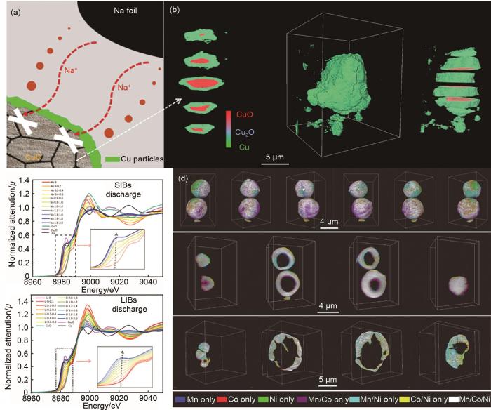
(a) passivation mechanism of “inactive” core of CuO particle; (b) sliced view of unreactive core CuO; (c) operando XANES spectra of CuO electrodes in SIBs and LIBs; (d) 3D elemental association maps of LiNi0.4Mn0.4Co0.2O2 particle[57]
ZUBI G, DUFO-LÓPEZ R, CARVALHO M, et al. The lithium-ion battery: State of the art and future perspectives[J]. Renewable and Sustainable Energy Reviews, 2018, 89: 292-308.
AN H W, LIU Q S, AN J L, et al. Coupling two-dimensional fillers with polymer chains in solid polymer electrolyte for room-temperature dendrite-free lithium-metal batteries[J]. Energy Storage Materials, 2021, 43: 358-364.
DONG L W, LIU J P, CHEN D J, et al. Suppression of polysulfide dissolution and shuttling with glutamate electrolyte for lithium sulfur batteries[J]. ACS Nano, 2019, 13(12): 14172-14181.
LI X, GONG Z L. Poly(ethylene oxide) based polymer electrolytes for all-solid-state Li-S batteries[J]. Journal of Electrochemistry, 2020, 26(3): 338-346.
PAN X N, LIU L L, WANG Z P, et al. The interaction of ternary components of ionic liquid gel polymer electrolytes for lithium metal batteries[J]. Journal of Electrochemistry, 2020, 26(3): 406-412.
MA Y L, ZHOU Z X, LI C J, et al. Enabling reliable lithium metal batteries by a bifunctional anionic electrolyte additive[J]. Energy Storage Materials, 2018, 11: 197-204.
LOU S F, YU Z J, LIU Q S, et al. Multi-scale imaging of solid-state battery interfaces: From atomic scale to macroscopic scale[J]. Chem, 2020, 6(9): 2199-2218.
TANG F C, WU Z B, YANG C, et al. Synchrotron X-ray tomography for rechargeable battery research: Fundamentals, setups and applications[J]. Small Methods, 2021, 5(9): doi: 10.1002/smtd. 202100557.
LI T, YANG H X. Electrochemical conversion reactions and their applications for rechargeable batteries[J]. Journal of Electrochemistry, 2015, 21(2): 115-122.
WANG J J, CHEN-WIEGART Y C K, WANG J. In situ three-dimensional synchrotron X-ray nanotomography of the (de)lithiation processes in tin anodes[J]. Angewandte Chemie International Edition, 2014, 53(17): 4460-4464.
WANG L G, WANG J J, GUO F M, et al. Understanding the initial irreversibility of metal sulfides for sodium-ion batteries via operando techniques[J]. Nano Energy, 2018, 43: 184-191.
SONG Y J, SUN X, REN L P, et al. Synchrotron X-ray characterization of metal-air batteries[J]. Journal of Electrochemistry, 2021, doi: 10.13208/j.electrochem.210846.
LOU S F, SUN N, ZHANG F, et al. Tracking battery dynamics by operando synchrotron X-ray imaging: Operation from liquid electrolytes to solid-state electrolytes[J]. Accounts of Materials Research, 2021, 2(12): 1177-1189.
HOU M Y, BAO H L, WANG K, et al. Electrochemical and in situ X-ray absorption fine structure study of Li-rich cathode materials[J]. Journal of Electrochemistry, 2016, 22(3): 288-298.
LOU S F, CHENG X Q, GAO J L, et al. Pseudocapacitive Li+ intercalation in porous Ti2Nb10O29 nanospheres enables ultra-fast lithium storage[J]. Energy Storage Materials, 2018, 11: 57-66.
WANG L G, WANG J J, ZUO P J. Probing battery electrochemistry with in operando synchrotron X-ray imaging techniques[J]. Small Methods, 2018, 2(8): doi: 10.1002/smtd.201700293.
SHEN F Y, DIXIT M B, XIAO X H, et al. Effect of pore connectivity on Li dendrite propagation within LLZO electrolytes observed with synchrotron X-ray tomography[J]. ACS Energy Letters, 2018, 3(4): 1056-1061.
WANG J J, CHEN-WIEGART Y C K, WANG J. In operando tracking phase transformation evolution of lithium iron phosphate with hard X-ray microscopy[J]. Nature Communications, 2014, 5: doi: 10.1038/ncomms5570.
ZHANG F, LOU S, LI S, et al. Surface regulation enables high stability of single-crystal lithium-ion cathodes at high voltage[J]. Nature Communications, 2020, 11: doi: 10.1038/S41467-020-16824-2.
NELSON WEKER J, TONEY M F. Emerging in situ and operando nanoscale X-ray imaging techniques for energy storage materials[J]. Advanced Functional Materials, 2015, 25(11): 1622-1637.
BEHRENS S, KAPPLER A, OBST M. Linking environmental processes to the in situ functioning of microorganisms by high-resolution secondary ion mass spectrometry (NanoSIMS) and scanning transmission X-ray microscopy (STXM)[J]. Environmental Microbiology, 2012, 14(11): 2851-2869.
LIM J, LI Y Y, ALSEM D H, et al. Origin and hysteresis of lithium compositional spatiodynamics within battery primary particles[J]. Science, 2016, 353(6299): 566-571.
LIU J, WANG B Q, BANIS M N, et al. Investigation of amorphous to crystalline phase transition of sodium titanate by X-ray absorption spectroscopy and scanning transmission X-ray microscopy[J]. Canadian Journal of Chemistry, 2017, 95(11): 1163-1169.
DE SMIT E, SWART I, CREEMER J F, et al. Nanoscale chemical imaging of a working catalyst by scanning transmission X-ray microscopy[J]. Nature, 2008, 456(7219): 222-225.
LOU S F, ZHANG F, FU C K, et al. Interface issues and challenges in all-solid-state batteries: Lithium, sodium, and beyond[J]. Advanced Materials, 2021, 33(6): doi: 10.1002/adma.202000721.
WANG J J, CHEN-WIEGART Y C, WANG J. In situ chemical mapping of a lithium-ion battery using full-field hard X-ray spectroscopic imaging[J]. Chemical Communications, 2013, 49(58): 6480-6482.
ZHAO Y, ZHENG K, SUN X L. Addressing interfacial issues in liquid-based and solid-state batteries by atomic and molecular layer deposition[J]. Joule, 2018, 2(12): 2583-2604.
LOU S F, LIU Q S, ZHANG F, et al. Insights into interfacial effect and local lithium-ion transport in polycrystalline cathodes of solid-state batteries[J]. Nature Communications, 2020, 11: doi: 10. 1038/S41467-020-19528-9.
DENG J J, VINE D J, CHEN S, et al. Simultaneous cryo X-ray ptychographic and fluorescence microscopy of green algae[J]. Proceedings of the National Academy of Sciences of the United States of America, 2015, 112(8): 2314-2319.
YANG Y, XU R, ZHANG K, et al. Quantification of heterogeneous degradation in Li-ion batteries[J]. Advanced Energy Materials, 2019, 9(25): doi: 10.1002/aenm.201900674.
XU Z R, RAHMAN M M, MU L Q, et al. Chemomechanical behaviors of layered cathode materials in alkali metal ion batteries[J]. Journal of Materials Chemistry A, 2018, 6(44): 21859-21884.
BANIS M N, YADEGARI H, SUN Q, et al. Revealing the charge/discharge mechanism of Na-O2 cells by in situ soft X-ray absorption spectroscopy[J]. Energy & Environmental Science, 2018, 11(8): 2073-2077.
HAIBEL A, MANKE I, MELZER A, et al. In situ microtomographic monitoring of discharging processes in alkaline cells[J]. Journal of the Electrochemical Society, 2010, 157(4): doi: 10.1149/1.3294566.
SHEARING P R, HOWARD L E, JØRGENSEN P S, et al. Characterization of the 3-dimensional microstructure of a graphite negative electrode from a Li-ion battery[J]. Electrochemistry Communications, 2010, 12(3): 374-377.
EASTWOOD D S, BRADLEY R S, TARIQ F, et al. The application of phase contrast X-ray techniques for imaging Li-ion battery electrodes[J]. Nuclear Instruments and Methods in Physics Research Section B: Beam Interactions with Materials and Atoms, 2014, 324: 118-123.
WALDMANN T, ITURRONDOBEITIA A, KASPER M, et al. Post-mortem analysis of aged lithium-ion batteries: Disassembly methodology and physico-chemical analysis techniques[J]. Journal of the Electrochemical Society, 2016, 163(10): A2149-A2164.
COOPER S J, EASTWOOD D S, GELB J, et al. Image based modelling of microstructural heterogeneity in LiFePO4 electrodes for Li-ion batteries[J]. Journal of Power Sources, 2014, 247: 1033-1039.
EBNER M, CHUNG D W, GARCíA R E, et al. Tortuosity anisotropy in Lithium-ion battery electrodes[J]. Advanced Energy Materials, 2014, 4(5): doi: 10.1002/aenm.201301278.
KEHRWALD D, SHEARING P R, BRANDON N P, et al. Local tortuosity inhomogeneities in a lithium battery composite electrode[J]. Journal of the Electrochemical Society, 2011, 158(12): doi: 10. 1149/2.079112jes.
FINEGAN D P, SCHEEL M, ROBINSON J B, et al. In-operando high-speed tomography of lithium-ion batteries during thermal runaway[J]. Nature Communications, 2015, 6: doi: 10.1038/ncomms7924.
QIAN G N, MONACO F, MENG D C, et al. The role of structural defects in commercial lithium-ion batteries[J]. Cell Reports Physical Science, 2021, 2(9): doi: 10.1016/j.xcrp.2021.100554.
BESLI M, XIA S H, KUPPAN S, et al. Mesoscale chemomechanical interplay of the LiNi0.8Co0.15Al0.05O2 cathode in solid-state polymer batteries[J]. Chemistry of Materials, 2019, 31(2): 491-501.
EBNER M, MARONE F, STAMPANONI M, et al. Visualization and quantification of electrochemical and mechanical degradation in Li ion batteries[J]. Science, 2013, 342(6159): 716-720.
LI J Z, LI S F, ZHANG Y X, et al. Multiphase, multiscale chemomechanics at extreme low temperatures: Battery electrodes for operation in a wide temperature range[J]. Advanced Energy Materials, 2021, 11(37): doi: 10.1002/aenm.202170143.
YU Z J, ZHANG X Y, FU C K, et al. Dendrites in solid-state batteries: Ion transport behavior, advanced characterization, and interface regulation[J]. Advanced Energy Materials, 2021, 11(18): doi: 10.1002/aenm.202003250.
LIU Q S, ZHU G, LI R H, et al. Fast lithium transport kinetics regulated by low energy-barrier LixMnO2 for long-life lithium metal batteries[J]. Energy Storage Materials, 2021, 41: 1-7.
EASTWOOD D S, BAYLEY P M, CHANG H J, et al. Three-dimensional characterization of electrodeposited lithium microstructures using synchrotron X-ray phase contrast imaging[J]. Chemical Communications, 2015, 51(2): 266-268.
HARRY K J, HALLINAN D T, PARKINSON D Y, et al. Detection of subsurface structures underneath dendrites formed on cycled lithium metal electrodes[J]. Nature Materials, 2014, 13(1): 69-73.
DIXIT M B, REGALA M, SHEN F Y, et al. Tortuosity effects in garnet-type Li7La3Zr2O12 solid electrolytes[J]. ACS Applied Materials & Interfaces, 2019, 11(2): 2022-2030.
KAN W H, WEI C X, CHEN D C, et al. Evolution of local structural ordering and chemical distribution upon delithiation of a rock salt-structured Li1.3Ta0.3Mn0.4O2 cathode[J]. Advanced Functional Materials, 2019, 29(17): doi: 10.1002/adfm.201808294.
SUN N, LIU Q S, CAO Y, et al. Anisotropically electrochemical-mechanical evolution in solid-state batteries and interfacial tailored strategy[J]. Angewandte Chemie International Edition, 2019, 58(51): 18647-18653.
YU Z J, WANG J J, WANG L G, et al. Unraveling the origins of the "unreactive core" in conversion electrodes to trigger high sodium-ion electrochemistry[J]. ACS Energy Letters, 2019, 4(8): 2007-2012.
LIN F, NORDLUND D, LI Y Y, et al. Metal segregation in hierarchically structured cathode materials for high-energy lithium batteries[J]. Nature Energy, 2016, 1: doi: 10.1038/nenergy.2015.4.
WANG J J, KAREN CHEN-WIEGART Y C, ENG C, et al. Visualization of anisotropic-isotropic phase transformation dynamics in battery electrode particles[J]. Nature Communications, 2016, 7: doi: 10.1038/ncomms12372.
YI T F, LI X Y, LIU H P, et al. Recent developments in the doping and surface modification of LiFePO4 as cathode material for power lithium ion battery[J]. Ionics, 2012, 18(6): 529-539.
LU X K, BERTEI A, FINEGAN D P, et al. 3D microstructure design of lithium-ion battery electrodes assisted by X-ray nano-computed tomography and modelling[J]. Nature Communications, 2020, 11: doi: 10.1038/S41467-020-15811-X.
ZHANG K, REN F, WANG X L, et al. Finding a needle in the haystack: Identification of functionally important minority phases in an operating battery[J]. Nano Letters, 2017, 17(12): 7782-7788.
QIAN G N, ZHANG J, CHU S Q, et al. Understanding the mesoscale degradation in nickel-rich cathode materials through machine-learning-revealed strain-redox decoupling[J]. ACS Energy Letters, 2021, 6(2): 687-693.
(a) working principles and data processing of TXM-XANES experiment on CuO anode<sup>[<xref ref-type="bibr" rid="R30">30</xref>]</sup>; (b) <i>operando</i> 2D spectroscopy imaging for characterization of single crystal LiNi<sub>0.6</sub>Co<sub>0.2</sub>Mn<sub>0.2</sub>O<sub>2</sub> particles and corresponding charged state distribution statistics<sup>[<xref ref-type="bibr" rid="R22">22</xref>]</sup>Fig. 2
... [22];(c) O元素和C元素的X射线荧光成像[37](a) X-rayfluorescence and ptychographic images of pristine; (b) 200th cycled NCM particles to show element distribution and morphology<sup>[<xref ref-type="bibr" rid="R22">22</xref>]</sup>; (c) X-rayfluorescence and ptychographic images of O and C<sup>[<xref ref-type="bibr" rid="R37">37</xref>]</sup>Fig. 4<strong>2.2</strong> 同步辐射三维成像
... [30];(b) 原位X射线二维谱学成像对于单晶LiNi0.6Co0.2Mn0.2O2 颗粒的表征以及对应的荷电态分布统计[22](a) working principles and data processing of TXM-XANES experiment on CuO anode<sup>[<xref ref-type="bibr" rid="R30">30</xref>]</sup>; (b) <i>operando</i> 2D spectroscopy imaging for characterization of single crystal LiNi<sub>0.6</sub>Co<sub>0.2</sub>Mn<sub>0.2</sub>O<sub>2</sub> particles and corresponding charged state distribution statistics<sup>[<xref ref-type="bibr" rid="R22">22</xref>]</sup>Fig. 2
... [30]; (b) operando 2D spectroscopy imaging for characterization of single crystal LiNi0.6Co0.2Mn0.2O2 particles and corresponding charged state distribution statistics[22]Fig. 2
... [33](a) solid polymer electrolyte cell; (b) schematic diagram of internal structure of solid-state battery model; (c) TXM-XANES mapping of single cathode particle as a function of charging time in ASSLBs; (d) corresponding schematic diagram to expound unique solid-state electrochemistry<sup>[<xref ref-type="bibr" rid="R33">33</xref>]</sup>Fig. 3
... [37](a) X-rayfluorescence and ptychographic images of pristine; (b) 200th cycled NCM particles to show element distribution and morphology<sup>[<xref ref-type="bibr" rid="R22">22</xref>]</sup>; (c) X-rayfluorescence and ptychographic images of O and C<sup>[<xref ref-type="bibr" rid="R37">37</xref>]</sup>Fig. 4<strong>2.2</strong> 同步辐射三维成像
... [45];(b) 从18650柱状锂离子电池收集缺陷区域的实验过程[46];(c) 带有4个切片图像的NCA颗粒的3D重构图像[47](a) 3D reconstructions of commercial 18650 NMC batteries<sup>[<xref ref-type="bibr" rid="R45">45</xref>]</sup>; (b) schematic illustration of experimental process for harvesting defective regions of interest from 18650-type Li-ion battery<sup>[<xref ref-type="bibr" rid="R46">46</xref>]</sup>; (c) 3D rendering of nanotomographic data of particle collected with four virtual slices<sup>[<xref ref-type="bibr" rid="R47">47</xref>]</sup>Fig. 5
除了正极之外,同步辐射断层扫描技术也被应用于锂电池负极的研究.在锂电解质界面,人们普遍认为具有高弹性模量的固体电解质可以有效阻挡锂金属枝晶的生长[50].然而,近期的研究表明锂离子仍能渗透固体电解质,最终使电池的循环稳定性和运行安全性恶化.枝晶引起的短路是开发高能量密度固态电池的最大瓶颈之一.虽然锂枝晶在固态电解质中生长的行为和机理还不完全清楚,但已证明紧密接触和均匀电流密度的固-固界面是均匀沉积锂的关键.在此基础上,本课题组[51]构建了三维Li x MnO2(LMO)电极骨架作为锂离子的快速输运通道,实现了Li的无枝晶溶解/沉积.利用同步加速器高分辨率X射线断层显微镜定量分析了电极的化学成分和孔隙率[图6(a)、(b)].研究表明,LMO在电极内部的均匀分布可以形成均衡的离子输运网络,促进空间锂离子通量的均匀分布.在锂沉积后,发现空隙被Li金属完全填充,循环电极呈现密集的锂沉积.基于同步纳米CT数据的连通性网络模型也显示离子通量传输距离均匀且较短,进一步说明快速的锂离子传输是均匀镀锂的关键.与重元素相比,碳和锂等原子序数较低的元素对硬X射线的衰减较弱,导致它们与电解质填充的孔隙空间的对比度较低.随着相位对比技术的兴起,X射线显微断层扫描技术可以增强不同弱衰减材料(如碳和锂)之间的对比度.Shearing等[52]成功实现了锂枝晶的同轴相位对比成像,实现了衰减和折射效应与衰减相似材料之间的图像界面解耦[图6(c)].相似地,图6(d)展示了X射线断层扫描技术对聚合物电解质/锂金属之间界面的研究[53]. ...
... [45]; (b) schematic illustration of experimental process for harvesting defective regions of interest from 18650-type Li-ion battery[46]; (c) 3D rendering of nanotomographic data of particle collected with four virtual slices[47]Fig. 5
除了正极之外,同步辐射断层扫描技术也被应用于锂电池负极的研究.在锂电解质界面,人们普遍认为具有高弹性模量的固体电解质可以有效阻挡锂金属枝晶的生长[50].然而,近期的研究表明锂离子仍能渗透固体电解质,最终使电池的循环稳定性和运行安全性恶化.枝晶引起的短路是开发高能量密度固态电池的最大瓶颈之一.虽然锂枝晶在固态电解质中生长的行为和机理还不完全清楚,但已证明紧密接触和均匀电流密度的固-固界面是均匀沉积锂的关键.在此基础上,本课题组[51]构建了三维Li x MnO2(LMO)电极骨架作为锂离子的快速输运通道,实现了Li的无枝晶溶解/沉积.利用同步加速器高分辨率X射线断层显微镜定量分析了电极的化学成分和孔隙率[图6(a)、(b)].研究表明,LMO在电极内部的均匀分布可以形成均衡的离子输运网络,促进空间锂离子通量的均匀分布.在锂沉积后,发现空隙被Li金属完全填充,循环电极呈现密集的锂沉积.基于同步纳米CT数据的连通性网络模型也显示离子通量传输距离均匀且较短,进一步说明快速的锂离子传输是均匀镀锂的关键.与重元素相比,碳和锂等原子序数较低的元素对硬X射线的衰减较弱,导致它们与电解质填充的孔隙空间的对比度较低.随着相位对比技术的兴起,X射线显微断层扫描技术可以增强不同弱衰减材料(如碳和锂)之间的对比度.Shearing等[52]成功实现了锂枝晶的同轴相位对比成像,实现了衰减和折射效应与衰减相似材料之间的图像界面解耦[图6(c)].相似地,图6(d)展示了X射线断层扫描技术对聚合物电解质/锂金属之间界面的研究[53]. ...
... [46];(c) 带有4个切片图像的NCA颗粒的3D重构图像[47](a) 3D reconstructions of commercial 18650 NMC batteries<sup>[<xref ref-type="bibr" rid="R45">45</xref>]</sup>; (b) schematic illustration of experimental process for harvesting defective regions of interest from 18650-type Li-ion battery<sup>[<xref ref-type="bibr" rid="R46">46</xref>]</sup>; (c) 3D rendering of nanotomographic data of particle collected with four virtual slices<sup>[<xref ref-type="bibr" rid="R47">47</xref>]</sup>Fig. 5
除了正极之外,同步辐射断层扫描技术也被应用于锂电池负极的研究.在锂电解质界面,人们普遍认为具有高弹性模量的固体电解质可以有效阻挡锂金属枝晶的生长[50].然而,近期的研究表明锂离子仍能渗透固体电解质,最终使电池的循环稳定性和运行安全性恶化.枝晶引起的短路是开发高能量密度固态电池的最大瓶颈之一.虽然锂枝晶在固态电解质中生长的行为和机理还不完全清楚,但已证明紧密接触和均匀电流密度的固-固界面是均匀沉积锂的关键.在此基础上,本课题组[51]构建了三维Li x MnO2(LMO)电极骨架作为锂离子的快速输运通道,实现了Li的无枝晶溶解/沉积.利用同步加速器高分辨率X射线断层显微镜定量分析了电极的化学成分和孔隙率[图6(a)、(b)].研究表明,LMO在电极内部的均匀分布可以形成均衡的离子输运网络,促进空间锂离子通量的均匀分布.在锂沉积后,发现空隙被Li金属完全填充,循环电极呈现密集的锂沉积.基于同步纳米CT数据的连通性网络模型也显示离子通量传输距离均匀且较短,进一步说明快速的锂离子传输是均匀镀锂的关键.与重元素相比,碳和锂等原子序数较低的元素对硬X射线的衰减较弱,导致它们与电解质填充的孔隙空间的对比度较低.随着相位对比技术的兴起,X射线显微断层扫描技术可以增强不同弱衰减材料(如碳和锂)之间的对比度.Shearing等[52]成功实现了锂枝晶的同轴相位对比成像,实现了衰减和折射效应与衰减相似材料之间的图像界面解耦[图6(c)].相似地,图6(d)展示了X射线断层扫描技术对聚合物电解质/锂金属之间界面的研究[53]. ...
... [46]; (c) 3D rendering of nanotomographic data of particle collected with four virtual slices[47]Fig. 5
除了正极之外,同步辐射断层扫描技术也被应用于锂电池负极的研究.在锂电解质界面,人们普遍认为具有高弹性模量的固体电解质可以有效阻挡锂金属枝晶的生长[50].然而,近期的研究表明锂离子仍能渗透固体电解质,最终使电池的循环稳定性和运行安全性恶化.枝晶引起的短路是开发高能量密度固态电池的最大瓶颈之一.虽然锂枝晶在固态电解质中生长的行为和机理还不完全清楚,但已证明紧密接触和均匀电流密度的固-固界面是均匀沉积锂的关键.在此基础上,本课题组[51]构建了三维Li x MnO2(LMO)电极骨架作为锂离子的快速输运通道,实现了Li的无枝晶溶解/沉积.利用同步加速器高分辨率X射线断层显微镜定量分析了电极的化学成分和孔隙率[图6(a)、(b)].研究表明,LMO在电极内部的均匀分布可以形成均衡的离子输运网络,促进空间锂离子通量的均匀分布.在锂沉积后,发现空隙被Li金属完全填充,循环电极呈现密集的锂沉积.基于同步纳米CT数据的连通性网络模型也显示离子通量传输距离均匀且较短,进一步说明快速的锂离子传输是均匀镀锂的关键.与重元素相比,碳和锂等原子序数较低的元素对硬X射线的衰减较弱,导致它们与电解质填充的孔隙空间的对比度较低.随着相位对比技术的兴起,X射线显微断层扫描技术可以增强不同弱衰减材料(如碳和锂)之间的对比度.Shearing等[52]成功实现了锂枝晶的同轴相位对比成像,实现了衰减和折射效应与衰减相似材料之间的图像界面解耦[图6(c)].相似地,图6(d)展示了X射线断层扫描技术对聚合物电解质/锂金属之间界面的研究[53]. ...
... [47](a) 3D reconstructions of commercial 18650 NMC batteries<sup>[<xref ref-type="bibr" rid="R45">45</xref>]</sup>; (b) schematic illustration of experimental process for harvesting defective regions of interest from 18650-type Li-ion battery<sup>[<xref ref-type="bibr" rid="R46">46</xref>]</sup>; (c) 3D rendering of nanotomographic data of particle collected with four virtual slices<sup>[<xref ref-type="bibr" rid="R47">47</xref>]</sup>Fig. 5
除了正极之外,同步辐射断层扫描技术也被应用于锂电池负极的研究.在锂电解质界面,人们普遍认为具有高弹性模量的固体电解质可以有效阻挡锂金属枝晶的生长[50].然而,近期的研究表明锂离子仍能渗透固体电解质,最终使电池的循环稳定性和运行安全性恶化.枝晶引起的短路是开发高能量密度固态电池的最大瓶颈之一.虽然锂枝晶在固态电解质中生长的行为和机理还不完全清楚,但已证明紧密接触和均匀电流密度的固-固界面是均匀沉积锂的关键.在此基础上,本课题组[51]构建了三维Li x MnO2(LMO)电极骨架作为锂离子的快速输运通道,实现了Li的无枝晶溶解/沉积.利用同步加速器高分辨率X射线断层显微镜定量分析了电极的化学成分和孔隙率[图6(a)、(b)].研究表明,LMO在电极内部的均匀分布可以形成均衡的离子输运网络,促进空间锂离子通量的均匀分布.在锂沉积后,发现空隙被Li金属完全填充,循环电极呈现密集的锂沉积.基于同步纳米CT数据的连通性网络模型也显示离子通量传输距离均匀且较短,进一步说明快速的锂离子传输是均匀镀锂的关键.与重元素相比,碳和锂等原子序数较低的元素对硬X射线的衰减较弱,导致它们与电解质填充的孔隙空间的对比度较低.随着相位对比技术的兴起,X射线显微断层扫描技术可以增强不同弱衰减材料(如碳和锂)之间的对比度.Shearing等[52]成功实现了锂枝晶的同轴相位对比成像,实现了衰减和折射效应与衰减相似材料之间的图像界面解耦[图6(c)].相似地,图6(d)展示了X射线断层扫描技术对聚合物电解质/锂金属之间界面的研究[53]. ...
... [47]Fig. 5
除了正极之外,同步辐射断层扫描技术也被应用于锂电池负极的研究.在锂电解质界面,人们普遍认为具有高弹性模量的固体电解质可以有效阻挡锂金属枝晶的生长[50].然而,近期的研究表明锂离子仍能渗透固体电解质,最终使电池的循环稳定性和运行安全性恶化.枝晶引起的短路是开发高能量密度固态电池的最大瓶颈之一.虽然锂枝晶在固态电解质中生长的行为和机理还不完全清楚,但已证明紧密接触和均匀电流密度的固-固界面是均匀沉积锂的关键.在此基础上,本课题组[51]构建了三维Li x MnO2(LMO)电极骨架作为锂离子的快速输运通道,实现了Li的无枝晶溶解/沉积.利用同步加速器高分辨率X射线断层显微镜定量分析了电极的化学成分和孔隙率[图6(a)、(b)].研究表明,LMO在电极内部的均匀分布可以形成均衡的离子输运网络,促进空间锂离子通量的均匀分布.在锂沉积后,发现空隙被Li金属完全填充,循环电极呈现密集的锂沉积.基于同步纳米CT数据的连通性网络模型也显示离子通量传输距离均匀且较短,进一步说明快速的锂离子传输是均匀镀锂的关键.与重元素相比,碳和锂等原子序数较低的元素对硬X射线的衰减较弱,导致它们与电解质填充的孔隙空间的对比度较低.随着相位对比技术的兴起,X射线显微断层扫描技术可以增强不同弱衰减材料(如碳和锂)之间的对比度.Shearing等[52]成功实现了锂枝晶的同轴相位对比成像,实现了衰减和折射效应与衰减相似材料之间的图像界面解耦[图6(c)].相似地,图6(d)展示了X射线断层扫描技术对聚合物电解质/锂金属之间界面的研究[53]. ...
... 除了正极之外,同步辐射断层扫描技术也被应用于锂电池负极的研究.在锂电解质界面,人们普遍认为具有高弹性模量的固体电解质可以有效阻挡锂金属枝晶的生长[50].然而,近期的研究表明锂离子仍能渗透固体电解质,最终使电池的循环稳定性和运行安全性恶化.枝晶引起的短路是开发高能量密度固态电池的最大瓶颈之一.虽然锂枝晶在固态电解质中生长的行为和机理还不完全清楚,但已证明紧密接触和均匀电流密度的固-固界面是均匀沉积锂的关键.在此基础上,本课题组[51]构建了三维Li x MnO2(LMO)电极骨架作为锂离子的快速输运通道,实现了Li的无枝晶溶解/沉积.利用同步加速器高分辨率X射线断层显微镜定量分析了电极的化学成分和孔隙率[图6(a)、(b)].研究表明,LMO在电极内部的均匀分布可以形成均衡的离子输运网络,促进空间锂离子通量的均匀分布.在锂沉积后,发现空隙被Li金属完全填充,循环电极呈现密集的锂沉积.基于同步纳米CT数据的连通性网络模型也显示离子通量传输距离均匀且较短,进一步说明快速的锂离子传输是均匀镀锂的关键.与重元素相比,碳和锂等原子序数较低的元素对硬X射线的衰减较弱,导致它们与电解质填充的孔隙空间的对比度较低.随着相位对比技术的兴起,X射线显微断层扫描技术可以增强不同弱衰减材料(如碳和锂)之间的对比度.Shearing等[52]成功实现了锂枝晶的同轴相位对比成像,实现了衰减和折射效应与衰减相似材料之间的图像界面解耦[图6(c)].相似地,图6(d)展示了X射线断层扫描技术对聚合物电解质/锂金属之间界面的研究[53]. ...
... [50]; (c) 3D rendering of Li microstructures under constant current (top), and higher density microstructures following plating and stripping (bottom); (d) Li dendrite growth with charge passed[52]Fig. 6
... 除了正极之外,同步辐射断层扫描技术也被应用于锂电池负极的研究.在锂电解质界面,人们普遍认为具有高弹性模量的固体电解质可以有效阻挡锂金属枝晶的生长[50].然而,近期的研究表明锂离子仍能渗透固体电解质,最终使电池的循环稳定性和运行安全性恶化.枝晶引起的短路是开发高能量密度固态电池的最大瓶颈之一.虽然锂枝晶在固态电解质中生长的行为和机理还不完全清楚,但已证明紧密接触和均匀电流密度的固-固界面是均匀沉积锂的关键.在此基础上,本课题组[51]构建了三维Li x MnO2(LMO)电极骨架作为锂离子的快速输运通道,实现了Li的无枝晶溶解/沉积.利用同步加速器高分辨率X射线断层显微镜定量分析了电极的化学成分和孔隙率[图6(a)、(b)].研究表明,LMO在电极内部的均匀分布可以形成均衡的离子输运网络,促进空间锂离子通量的均匀分布.在锂沉积后,发现空隙被Li金属完全填充,循环电极呈现密集的锂沉积.基于同步纳米CT数据的连通性网络模型也显示离子通量传输距离均匀且较短,进一步说明快速的锂离子传输是均匀镀锂的关键.与重元素相比,碳和锂等原子序数较低的元素对硬X射线的衰减较弱,导致它们与电解质填充的孔隙空间的对比度较低.随着相位对比技术的兴起,X射线显微断层扫描技术可以增强不同弱衰减材料(如碳和锂)之间的对比度.Shearing等[52]成功实现了锂枝晶的同轴相位对比成像,实现了衰减和折射效应与衰减相似材料之间的图像界面解耦[图6(c)].相似地,图6(d)展示了X射线断层扫描技术对聚合物电解质/锂金属之间界面的研究[53]. ...
... [51];(c) 恒流充电条件下的锂金属微观结构3D重构图(上),沉积/溶解后的高密度微观结构(下);(d) 通过X射线断层扫描技术观察到的锂枝晶生长过程[52](a) X-ray nanotomography and virtual slice of composite Li anode and cycled anode; (b) connectivity network of cycled composite Li electrode; intensity bar indicates Li<sup>+</sup> transport distance<sup>[<xref ref-type="bibr" rid="R50">50</xref>]</sup>; (c) 3D rendering of Li microstructures under constant current (top), and higher density microstructures following plating and stripping (bottom); (d) Li dendrite growth with charge passed<sup>[<xref ref-type="bibr" rid="R52">52</xref>]</sup>Fig. 6
... 除了正极之外,同步辐射断层扫描技术也被应用于锂电池负极的研究.在锂电解质界面,人们普遍认为具有高弹性模量的固体电解质可以有效阻挡锂金属枝晶的生长[50].然而,近期的研究表明锂离子仍能渗透固体电解质,最终使电池的循环稳定性和运行安全性恶化.枝晶引起的短路是开发高能量密度固态电池的最大瓶颈之一.虽然锂枝晶在固态电解质中生长的行为和机理还不完全清楚,但已证明紧密接触和均匀电流密度的固-固界面是均匀沉积锂的关键.在此基础上,本课题组[51]构建了三维Li x MnO2(LMO)电极骨架作为锂离子的快速输运通道,实现了Li的无枝晶溶解/沉积.利用同步加速器高分辨率X射线断层显微镜定量分析了电极的化学成分和孔隙率[图6(a)、(b)].研究表明,LMO在电极内部的均匀分布可以形成均衡的离子输运网络,促进空间锂离子通量的均匀分布.在锂沉积后,发现空隙被Li金属完全填充,循环电极呈现密集的锂沉积.基于同步纳米CT数据的连通性网络模型也显示离子通量传输距离均匀且较短,进一步说明快速的锂离子传输是均匀镀锂的关键.与重元素相比,碳和锂等原子序数较低的元素对硬X射线的衰减较弱,导致它们与电解质填充的孔隙空间的对比度较低.随着相位对比技术的兴起,X射线显微断层扫描技术可以增强不同弱衰减材料(如碳和锂)之间的对比度.Shearing等[52]成功实现了锂枝晶的同轴相位对比成像,实现了衰减和折射效应与衰减相似材料之间的图像界面解耦[图6(c)].相似地,图6(d)展示了X射线断层扫描技术对聚合物电解质/锂金属之间界面的研究[53]. ...
... [52](a) X-ray nanotomography and virtual slice of composite Li anode and cycled anode; (b) connectivity network of cycled composite Li electrode; intensity bar indicates Li<sup>+</sup> transport distance<sup>[<xref ref-type="bibr" rid="R50">50</xref>]</sup>; (c) 3D rendering of Li microstructures under constant current (top), and higher density microstructures following plating and stripping (bottom); (d) Li dendrite growth with charge passed<sup>[<xref ref-type="bibr" rid="R52">52</xref>]</sup>Fig. 6
... 除了正极之外,同步辐射断层扫描技术也被应用于锂电池负极的研究.在锂电解质界面,人们普遍认为具有高弹性模量的固体电解质可以有效阻挡锂金属枝晶的生长[50].然而,近期的研究表明锂离子仍能渗透固体电解质,最终使电池的循环稳定性和运行安全性恶化.枝晶引起的短路是开发高能量密度固态电池的最大瓶颈之一.虽然锂枝晶在固态电解质中生长的行为和机理还不完全清楚,但已证明紧密接触和均匀电流密度的固-固界面是均匀沉积锂的关键.在此基础上,本课题组[51]构建了三维Li x MnO2(LMO)电极骨架作为锂离子的快速输运通道,实现了Li的无枝晶溶解/沉积.利用同步加速器高分辨率X射线断层显微镜定量分析了电极的化学成分和孔隙率[图6(a)、(b)].研究表明,LMO在电极内部的均匀分布可以形成均衡的离子输运网络,促进空间锂离子通量的均匀分布.在锂沉积后,发现空隙被Li金属完全填充,循环电极呈现密集的锂沉积.基于同步纳米CT数据的连通性网络模型也显示离子通量传输距离均匀且较短,进一步说明快速的锂离子传输是均匀镀锂的关键.与重元素相比,碳和锂等原子序数较低的元素对硬X射线的衰减较弱,导致它们与电解质填充的孔隙空间的对比度较低.随着相位对比技术的兴起,X射线显微断层扫描技术可以增强不同弱衰减材料(如碳和锂)之间的对比度.Shearing等[52]成功实现了锂枝晶的同轴相位对比成像,实现了衰减和折射效应与衰减相似材料之间的图像界面解耦[图6(c)].相似地,图6(d)展示了X射线断层扫描技术对聚合物电解质/锂金属之间界面的研究[53]. ...
... [55];(d) FeS2 颗粒内部微观结构的剖视图以及所选截面的化学相映射、吸收光谱[56](a) 3D mapping of Mn of a Li <i><sub>x</sub></i> Ta<sub>0.3</sub>Mn<sub>0.4</sub>O<sub>2</sub> (LTMO) particle; (b) 3D morphology mapping of an LTMO particle<sup>[<xref ref-type="bibr" rid="R55">55</xref>]</sup>; (c) cross-sectional slides of FeS<sub>2</sub> particle along <i>X</i>-<i>Y</i>, <i>Y</i>-<i>Z</i> and <i>X</i>-<i>Z</i> plane; (d) internal microstructure of same particle with a cut-away view<sup>[<xref ref-type="bibr" rid="R56">56</xref>]</sup>Fig. 8
... [55]; (c) cross-sectional slides of FeS2 particle along X-Y, Y-Z and X-Z plane; (d) internal microstructure of same particle with a cut-away view[56]Fig. 8
(a) 3D mapping of Mn of a Li <i><sub>x</sub></i> Ta<sub>0.3</sub>Mn<sub>0.4</sub>O<sub>2</sub> (LTMO) particle; (b) 3D morphology mapping of an LTMO particle<sup>[<xref ref-type="bibr" rid="R55">55</xref>]</sup>; (c) cross-sectional slides of FeS<sub>2</sub> particle along <i>X</i>-<i>Y</i>, <i>Y</i>-<i>Z</i> and <i>X</i>-<i>Z</i> plane; (d) internal microstructure of same particle with a cut-away view<sup>[<xref ref-type="bibr" rid="R56">56</xref>]</sup>Fig. 8
... [57](a) passivation mechanism of “inactive” core of CuO particle; (b) sliced view of unreactive core CuO; (c) <i>operando</i> XANES spectra of CuO electrodes in SIBs and LIBs; (d) 3D elemental association maps of LiNi<sub>0.4</sub>Mn<sub>0.4</sub>Co<sub>0.2</sub>O<sub>2</sub> particle<sup>[<xref ref-type="bibr" rid="R57">57</xref>]</sup>Fig. 9<strong>2.4</strong> 五维成像技术
... [59](a) schematic of experimental setup of 5D transmission X-ray microscopy; (b) phase distribution as a function of charging time<sup>[<xref ref-type="bibr" rid="R59">59</xref>]</sup>Fig. 10


