Research progress in understanding of lithium storage behavior and reaction mechanism of electrode materials through in situ transmission electron microscopy
KE Chengzhi,1, XIAO Bensheng1, LI Miao1, LU Jingyu3, HE Yang4, ZHANG Li2, ZHANG Qiaobao,1
1.Department of Materials Science and Engineering, College of Materials, Xiamen University
2.College of Chemistry and Chemical Engineering, Xiamen University, Xiamen 361005, Fujian, China
3.School of Science, Harbin Institute of Technology (Shenzhen), Shenzhen 518055, Guangdong, China
4.Beijing Advanced Innovation Center for Materials Genome Engineering, University of Science and Technology Beijing, Beijing 100083, China
Transport, reaction, and storage of Li ions in bulk electrode materials leads to the dynamic evolution of their electronic and crystal structures, microstructures, chemical compositions, and physical properties, which are the important determinants in the electrochemical performance of Li ion batteries. It is extremely important to understand the fundamental physical and chemical properties of electrode materials at a nanometer or even atomic scale to determine their microstructure, morphology, phase, and chemical composition during an electrochemical process. The structure-activity relationship between the electrode materials and macroscopic electrochemical performance of a battery must be considered. These require clear, precise, and advanced in situ characterizations. Among existing in situ characterization techniques, in situ transmission electron microscopy (TEM) is one of the most representative and important methods to conduct these experiments. Its unique advantages include ultra-high spatial and temporal resolution and real-time, dynamic monitoring of the structure, morphology, phase and interface evolution of the electrode materials under the given working conditions. It can precisely evaluate the microscopic dynamic evolution behavior and the reaction mechanism of electrode materials, providing a microscopic basis and innovative ideas for the construction and performance regulation of high-performance electrode materials. In this study, we summarize important progress on in situ TEM investigations of the dynamic evolution and failure mechanism of key electrode materials in Li ion batteries during charge/discharge. These investigations include various cathode materials and high specific capacity anode materials, especially their dynamic evolutions in microstructure, chemical composition, and phases during an electrochemical process. Moreover, the current limitations of in situ TEM and future directions of in situ TEM investigation of secondary batteries are discussed.
Keywords:electrode materials
;
lithium ion storage behavior
;
lithium ion storage mechanisms
;
in situ transmission electron microscopy
;
lithium ion batteries
KE Chengzhi. Research progress in understanding of lithium storage behavior and reaction mechanism of electrode materials through in situ transmission electron microscopy[J]. Energy Storage Science and Technology, 2021, 10(4): 1219-1236
Fig. 1
(a) HAADF image of delithiated LiCoO2 cathode after coloring with GPA method, (b) and (c) are zoomed-in views of yellow, dashed-box area, (d) and (e) are zoomed-in images of pink, dashed-box area[37], (f) A HAADF-STEM image of a pristine thin film LCO piece, (g) A TEM image after lithiation acquired at a low magnification, (h)~(k) NBD patterns for different spots labeled in (g)[38]
Fig. 2
(a) Schematic diagram of in situ TEM setup with nanobattery, (b) A TEM image of FePO4 particle after its contact with Li/Li2O solid electrolyte, (c)~(i) Time-lapse TEM images of FePO4 particle during lithiation[42], (j)~(l) Microstructural evolution of a LFP nanowire during lithiation, (m)、(n) Magnified images of dotted red rectangle region in (j)、(k), respectively. Simulated TEM images of (o) LiFePO4 and (p) Li0.5FePO4[43]
Fig. 3
(a) Schematic of in situ TEM experimental setup; (b) low magnification HAADF-STEM images; (c),(d)HR-HAADF-STEM images of NMC before and after delithiation, respectively; (e) Phases extension and defects migration; (f)~(h) Magnified images of (c),(d)[44]
Fig. 4
(a) HAADF-STEM image of antiphase boundaries, (b)~(d) EELS spectrum of O, Mn, and Ni from (a), respectively, (e) HAADF-STEM image of migration-front area, (f)~(h) EELS spectrum of O, Mn, and Ni from (e), (i)~(k)ABF-STEM images of pristine and charged LNMO along [111], [110] and [100] direction, respectively[45]
Fig. 5
(a)~(l) Top-view SEM images of Si nanopillars along [100], [110] and [111] directions at different lithiation states; (m), (n) Statistical data of changes in cross-sectional dimensions and cross-sectional area along these three orientations[55]; (o), (p) Microstructural evolution of Si nanowire during lithiation; (q) 3D simulation plot of a progressively lithiated Si nanowire[56]
Fig. 6
(a)~(d) TEM images and corresponding electron diffraction patterns of a pristine silicon nanowire and a partially lithiated silicon nanowire; (e)~(g) Time-lapse images showing migration of ACI during lithiation; (h)~(j) TEM images and diagrams of lithiation process by lateral ledge flow in ACI and simultaneous peeling-off[57]
Fig. 7
(a)~(e) TEM images showing fast fracture of 620 nm Si nanoparticle during lithiation process. (f) Electron diffraction pattern of broken Si nanoparticle after lithiation, indicating formation of Li15Si4. (g)~(j) TEM images of a 80 nm Si nanoparticle without cracking during lithiation. (k) Statistics showing critical size of Si nanoparticles without cracking during lithiation process[58]
Fig. 8
(a) TEM image of a Cu/Si/Ge nanowire; (b) HRTEM image of Si/Ge bilayer and corresponding SAED pattern; (c) STEM image of Cu/Si/Ge nanowire and corresponding EDX spectra; (d), (e) Time-lapse TEM images of Cu/Si/Ge nanowire during lithiation; (f) Evolution of layer thickness in Si/Ge bilayer shell during lithiation[60]
Fig. 9
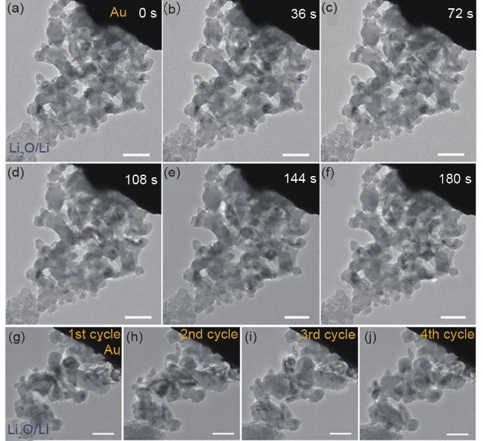
(a)~(f) Time-lapse TEM images of AMPSi@C electrode during lithiation. In situ TEM images of AMPSi@C (g) after 1st cycle, (h) after 2nd cycle, (i) after 3rd cycle and (j) after 4th cycle[65]
Fig. 10
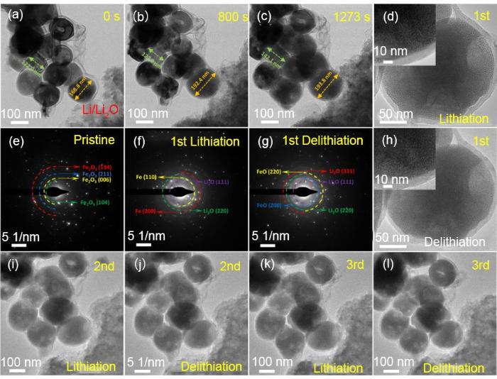
(a)~(c) Time-lapse TEM images of a yolk@shell Fe2O3@C electrode during first lithiation process; TEM images acquired after first lithiation (d), and first delithiation (h); SAED patterns before lithiation (e), after first lithiation (f) and after first delithiation (g); TEM images after second lithiation (i), after second delithiation (j), after third lithiation (k) and after third delithiation (l)[73]
Fig. 11
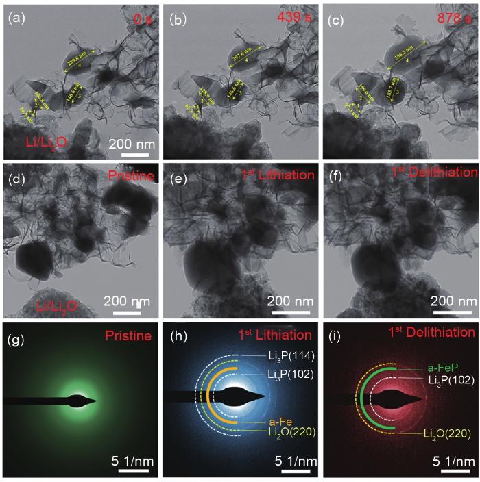
(a)~(c) Time-resolved TEM images of a FeP@CNs electrode during lithiation. In situ TEM images and corresponding SAED patterns of pristine FeP@CNs electrode (d)~(g), after first lithiation [(e), (h)] and first delithiation [(f), (i)][74]
缪平, 姚祯, LEMMON J, 等. 电池储能技术研究进展及展望[J]. 储能科学与技术, 2020, 9(3): 670-678.MIAO P, YAO Z, LEMMON J, et al. Current situations and prospects of energy storage batteries[J]. Energy Storage Science and Technology, 2020, 9(3): 670-678.
ZHONG S Y, SHI J, LUO W W, et al. First-principles insight into Li and Na ion storage in graphene oxide[J]. Chinese Physics B, 2019, 28(7): 591-597.
ZHAO L Z, WU H H, YANG C H, et al. Mechanistic origin of the high performance of Yolk@Shell Bi2S3@N-doped carbon nanowire electrodes[J]. ACS Nano, 2018, 12(12): 12597-12611.
ARMAND M, TARASCON J M. Building better batteries[J]. Nature, 2008, 451(7179): 652-657.
张桥保, 龚正良, 杨勇. 硫化物固态电解质材料界面及其表征的研究进展[J]. 物理学报, 2020, 69(22): 153-180.ZHANG Q B, GONG Z L, YANG Y. Advance in interface and characterizations of sulfide solid electrolyte materials[J]. Acta Physica Sinica, 2020, 69(22): 153-180.
岳昕阳, 马萃, 包戬, 等. 金属锂负极失效机制及其先进表征技术[J]. 物理化学学报, 2021, 37(2): 8-29.YUE X Y, MA C, BAO J, et al. Failure mechanisms of lithium metal anode and their advanced characterization technologies[J]. Acta Physico Chimica Sinica, 2021, 37(2): 8-29.
拱越, 谷林. 全固态电池中界面的结构演化和物质输运[J]. 物理学报, 2020, 69(22): 57-64.GONG Y, Gu L. Structural evolution and matter transportation of the interface in all-solid-state battery[J]. Acta Physica Sinica, 2020, 69(22): 57-64.
DUNN B, KAMATH H, TARASCON J M. Electrical energy storage for the grid: a battery of choices[J]. Science, 2011, 334(6058): 928-935.
GOODENOUGH J B, PARK K S. The Li-ion rechargeable battery: a perspective[J]. Journal of the American Chemical Society, 2013, 135(4): 1167-1176.
LARCHER D, TARASCON J M. Towards greener and more sustainable batteries for electrical energy storage[J]. Nature Chemistry, 2015, 7(1): 19-29.
XIANG Y X, LI X, CHENG Y Q, et al. Advanced characterization techniques for solid state lithium battery research[J]. Materials Today, 2020, 36: 139-157.
WANG Z Y, SANTHANAGOPALAN D, ZHANG W, et al. In situ STEM-EELS observation of nanoscale interfacial phenomena in all-solid-state batteries[J]. Nano Letters, 2016, 16(6): 3760-3766.
LIU X H, HUANG J Y. In situ TEM electrochemistry of anode materials in lithium ion batteries[J]. Energy & Environmental Science, 2011, 4(10): 3844-3860.
WU X Y, LI S M, YANG B, et al. In situ transmission electron microscopy studies of electrochemical reaction mechanisms in rechargeable batteries[J]. Electrochemical Energy Reviews, 2019, 2(3): 467-491.
CHENG Y, ZHANG L Q, ZHANG Q B, et al. Understanding all solid-state lithium batteries through in situ transmission electron microscopy[J]. Materials Today, 2020, 42: 137-161.
徐涛, 孙俊, 孙立涛. 原位动态电子显微学研究进展[J]. 物理学进展, 2012, 32(3): 115-134.XU T, SUN J, SUN L T. Progress in dynamic in situ electron microscopy[J]. Progress in Physics, 2012, 32(3): 115-134.
ZHANG C, FIRESTEIN K L, FERNANDO J F S, et al. Recent progress of in situ transmission electron microscopy for energy materials[J]. Advanced Materials, 2020, 32(18): e1904094.
HOLTZ M E, YU Y C, GUNCELER D, et al. Nanoscale imaging of lithium ion distribution during in situ operation of battery electrode and electrolyte[J]. Nano Letters, 2014, 14(3): 1453-1459.
LIU X H, LIU Y, KUSHIMA A, et al. In situ TEM experiments of electrochemical lithiation and delithiation of individual nanostructures[J]. Advanced Energy Materials, 2012, 2(7): 722-741.
MA X Y, LUO W, YAN M Y, et al. In situ characterization of electrochemical processes in one dimensional nanomaterials for energy storages devices[J]. Nano Energy, 2016, 24: 165-188.
赵一博, 刘蕙蕙, 陈松良, 等. 先进成像技术在全固态锂电池关键问题研究中的应用[J]. 电化学, 2019, 25(1): 17-30.ZHAO Y B, LIU H H, CHEN S L, et al. Applications of advanced imaging technologies for critical issues of all-solid-state lithium battery studies[J]. Journal of Electrochemistry, 2019, 25(1): 17-30.
李文俊, 褚赓, 彭佳悦, 等. 锂离子电池基础科学问题(Ⅻ)——表征方法[J]. 储能科学与技术, 2014, 3(6): 642-667.LI W J, CHU G, PENG J Y, et al. Fundamental scientific aspects of lithium batteries (Ⅻ)-Characterization techniques[J]. Energy Storage Science and Technology, 2014, 3(6): 642-667.
LEE H W, LI Y Z, CUI Y. Perspectives in in situ transmission electron microscopy studies on lithium battery electrodes[J]. Current Opinion in Chemical Engineering, 2016, 12: 37-43.
QI K, WEI J K, SUN M H, et al. Real-time observation of deep lithiation of tungsten oxide nanowires by in situ electron microscopy[J]. Angewandte Chemie International Edition, 2015, 54(50): 15222-15225.
苏庆梅, 杜高辉, 郭俊杰, 等. 原位透射电镜技术在电化学储能领域的研究进展[J]. 中国材料进展, 2020, 39(Z1): 559-575+557-558.SU Q M, DU G H, GUO J J, et al. Recent progress of in situ transmission electron microscopy on electrochemical energy storage[J]. Materials China, 2020, 39(Z1): 559-575+557-558.
YANG J, MUHAMMAD S, JO M R, et al. In situ analyses for ion storage materials[J]. Chemical Society Reviews, 2016, 45(20): 5717-5770.
LIU N, LU Z D, ZHAO J, et al. A pomegranate-inspired nanoscale design for large-volume-change lithium battery anodes[J]. Nature Nanotechnology, 2014, 9(3): 187-192.
拱越, 谷林. 锂离子电池材料的电子显微学分析方法[J]. 储能科学与技术, 2019, 8(6): 1260-1270.GONG Y, GU L. Transmission electron microscopy of lithium ion battery materials[J]. Energy Storage Science and Technology, 2019, 8(6): 1260-1270.
张利强, 唐永福, 刘秋男, 等. 原位透射电镜技术在电池领域的研究进展[J]. 储能科学与技术, 2019, 8(6): 1050-1061.ZHANG L Q, TANG Y F, LIU Q N, et al. Review of in situ transmission electron microscopy studies of battery materials[J]. Energy Storage Science and Technology, 2019, 8(6): 1050-1061.
YUAN Y F, AMINE K, LU J, et al. Understanding materials challenges for rechargeable ion batteries with in situ transmission electron microscopy[J]. Nature Communications, 2017, 8(1): 15806.
WANG Y, CAO G Z. Developments in nanostructured cathode materials for high-performance lithium-ion batteries[J]. Advanced Materials, 2008, 20(12): 2251-2269.
REIMERS J N, DAHN J R. Electrochemical and in situ X-ray diffraction studies of lithium intercalation in LixCoO2[J]. Journal of the Electrochemical Society, 1992, 139(8): 2091.
蓝兹炜, 张建茹, 李园园, 等. 基于锂离子电池正极材料的一元/二元复合正极材料研究进展[J]. 储能科学与技术, 2021, 10(1): 27-39.LAN Z W, ZHANG J R, LI Y Y, et al. Research progress of mono/binary composite cathode materials based on lithium-ion battery cathode materials[J]. Energy Storage Science and Technology, 2021, 10(1): 27-39.
GONG Y, ZHANG J N, JIANG L W, et al. In situ atomic-scale observation of electrochemical delithiation induced structure evolution of LiCoO2 cathode in a working all-solid-state battery[J]. Journal of the American Chemical Society, 2017, 139(12): 4274-4277.
李文俊, 郑杰允, 谷林, 等. 锂电池原位与非原位表征技术研究[J]. 电化学, 2015, 21(2): 99-114.LI W J, ZHENG J Y, GU L, et al. Researches on in-situ and ex-situ characterization techniques in lithium batteries[J]. Journal of Electrochemistry, 2015, 21(2): 99-114.
SHU J, SHUI M, HUANG F T, et al. A new look at lithium cobalt oxide in a broad voltage range for lithium-ion batteries[J]. Journal of Physical Chemistry C, 2010, 114(7): 3323-3328.
WEKER J N, WISE A M, LIM K, et al. Operando spectroscopic microscopy of LiCoO2 cathodes outside standard operating potentials[J]. Electrochimica Acta, 2017, 247: 977-982.
ZHU Y J, WANG J W, LIU Y, et al. In situ atomic-scale imaging of phase boundary migration in FePO4 microparticles during electrochemical lithiation[J]. Advanced Materials, 2013, 25(38): 5461-5466.
LI S, YAO Z P, ZHENG J M, et al. Direct observation of defect-aided structural evolution in a nickel-rich layered cathode[J]. Angewandte Chemie International Edition, 2020, 59(49): 22092-22099.
GONG Y, CHEN Y Y, ZHANG Q H, et al. Three-dimensional atomic-scale observation of structural evolution of cathode material in a working all-solid-state battery[J]. Nature Communications, 2018, 9(1): 3341.
麦立强, 陈丹丹, 赵康宁, 等. 纳米线电化学储能材料与器件[J]. 科学通报, 2013, 58(32): 3312-3327.MAI L Q, CHEN D D, ZHAO K N, et al. Nanowire device for electrochemical energy storage[J]. Chinese Science Bulletin, 2013, 58(32): 3312-3327.
余晨露, 田晓华, 郑瀚, 等. 高稳定性硅/硬碳复合负极在锂电负极中的应用[J]. 储能科学与技术, 2021, 10(1): 128-136.YU C L, TIAN X H, ZHANG H, et al. Research progress in high stability of silicon/hard carbon anodes for LIBs[J]. Energy Storage Science and Technology, 2021, 10(1): 128-136.
ZHENG Z, WU H H, CHEN H, et al. Fabrication and understanding of Cu3Si-Si@carbon@graphene nanocomposites as high-performance anodes for lithium-ion batteries[J]. Nanoscale, 2018, 10(47): 22203-22214.
HE Y, PIPER D M, GU M, et al. In situ transmission electron microscopy probing of native oxide and artificial layers on silicon nanoparticles for lithium ion batteries[J]. ACS Nano, 2014, 8(11): 11816-23.
GU M, LI Y, LI X L, et al. In situ TEM study of lithiation behavior of silicon nanoparticles attached to and embedded in a carbon matrix[J]. ACS Nano, 2012, 6(9): 8439-8447.
MCDOWELL M T, LEE S W, WANG C M, et al. The effect of metallic coatings and crystallinity on the volume expansion of silicon during electrochemical lithiation/delithiation[J]. Nano Energy, 2012, 1(3): 401-410.
GHASSEMI H, AU M, CHEN N, et al. In situ electrochemical lithiation/delithiation observation of individual amorphous Si nanorods[J]. ACS Nano, 2011, 5(10): 7805-7811.
LEE S W, MCDOWELL M T, CHOI J W, et al. Anomalous shape changes of silicon nanopillars by electrochemical lithiation[J]. Nano Letters, 2011, 11(7): 3034-3039.
LIU X H, WANG J W, HUANG S, et al. In situ atomic-scale imaging of electrochemical lithiation in silicon[J]. Nature Nanotechnology, 2012, 7(11): 749-56.
WANG J W, LIU X H, ZHAO K, et al. Sandwich-lithiation and longitudinal crack in amorphous silicon coated on carbon nanofibers[J]. ACS Nano, 2012, 6(10): 9158-67.
ZHANG Q B, CHEN H X, LUO L L, et al. Harnessing the concurrent reaction dynamics in active Si and Ge to achieve high performance lithium-ion batteries[J]. Energy & Environmental Science, 2018, 11(3): 669-681.
WANG H K, YANG X M, WU Q Z, et al. Encapsulating silica/antimony into porous electrospun carbon nanofibers with robust structure stability for high-efficiency lithium storage[J]. ACS Nano, 2018, 12(4): 3406-3416.
WANG C M, LI X L, WANG Z G, et al. In situ TEM investigation of congruent phase transition and structural evolution of nanostructured silicon/carbon anode for lithium ion batteries[J]. Nano Letters, 2012, 12(3): 1624-1632.
SON I H, HWAN PARK J, KWON S, et al. Silicon carbide-free graphene growth on silicon for lithium-ion battery with high volumetric energy density[J]. Nature Communications, 2015, 6: 7393.
LI Y Z, YAN K, LEE H W, et al. Growth of conformal graphene cages on micrometre-sized silicon particles as stable battery anodes[J]. Nature Energy, 2016, 1(2): 15029.
AN W L, GAO B, MEI S X, et al. Scalable synthesis of ant-nest-like bulk porous silicon for high-performance lithium-ion battery anodes[J]. Nature Communications, 2019, 10(1): 1447.
LU Z D, LIU N, LEE H W, et al. Nonfilling carbon coating of porous silicon micrometer-sized particles for high-performance lithium battery anodes[J]. ACS Nano, 2015, 9(3): 2540-2547.
LIU X H, HUANG S, PICRAUX S T, et al. Reversible nanopore formation in Ge nanowires during lithiation-delithiation cycling: An in situ transmission electron microscopy study[J]. Nano Letters, 2011, 11(9): 3991-3997.
BOEBINGER M G, YAREMA O, YAREMA M, et al. Spontaneous and reversible hollowing of alloy anode nanocrystals for stable battery cycling[J]. Nature Nanotechnology, 2020, 15(6): 475-481.
LI Y Y, OU C Z, ZHU J L, et al. Ultrahigh and durable volumetric lithium/sodium storage enabled by a highly dense graphene-encapsulated nitrogen-doped carbon@Sn compact monolith[J]. Nano Letters, 2020, 20(3): 2034-2046.
LIANG W T, HONG L, YANG H, et al. Nanovoid formation and annihilation in gallium nanodroplets under lithiation-delithiation cycling[J]. Nano Letters, 2013, 13(11): 5212-5217.
SU Q M, XIE D, ZHANG J, et al. In situ transmission electron microscopy observation of the conversion mechanism of Fe2O3/graphene anode during lithiation-delithiation processes[J]. ACS Nano, 2013, 7(10): 9115-9121.
YU W J, ZHANG L L, HOU P X, et al. High reversible lithium storage capacity and structural changes of Fe2O3 nanoparticles confined inside carbon nanotubes[J]. Advanced Energy Materials, 2016, 6(3): 1501755.
ZHENG Z M, LI P, HUANG J, et al. High performance columnar-like Fe2O3@carbon composite anode via yolk@shell structural design[J]. Journal of Energy Chemistry, 2020, 41: 126-134.
ZHENG Z M, WU H H, LIU H D, et al. Achieving fast and durable lithium storage through amorphous FeP nanoparticles encapsulated in ultrathin 3D P-doped porous carbon nanosheets[J]. ACS Nano, 2020, 14(8): 9545-9561.
HE K, ZHANG S, LI J, et al. Visualizing non-equilibrium lithiation of spinel oxide via in situ transmission electron microscopy[J]. Nature Communications, 2016, 7: 11441.
WANG X, TANG D M, LI H Q, et al. Revealing the conversion mechanism of CuO nanowires during lithiation-delithiation by in situ transmission electron microscopy[J]. Chemical Communications, 2012, 48(40): 4812-4814.
HE K, LIN F, ZHU Y Z, et al. Sodiation kinetics of metal oxide conversion electrodes: A comparative study with lithiation[J]. Nano Letters, 2015, 15(9): 5755-5763.
GREGORCZYK K E, LIU Y, SULLIVAN J P, et al. In situ transmission electron microscopy study of electrochemical lithiation and delithiation cycling of the conversion anode RuO2[J]. ACS Nano, 2013, 7(7): 6354-6360.
CHEN K J, CAO K, XING C C, et al. In-situ TEM study of the lithiation and delithiation of FeS nanosheets[J]. Journal of Alloys and Compounds, 2016, 688: 946-952.
SU Q M, XIE J, ZHANG J, et al. In situ transmission electron microscopy observation of electrochemical behavior of CoS2 in lithium-ion battery[J]. ACS Applied Materials & Interfaces, 2014, 6(4): 3016-3022.
HE Y, GU M, XIAO H Y, et al. Atomistic conversion reaction mechanism of WO3 in secondary ion batteries of Li, Na, and Ca[J]. Angewandte Chemie International Edition, 2016, 55(21): 6244-6247.
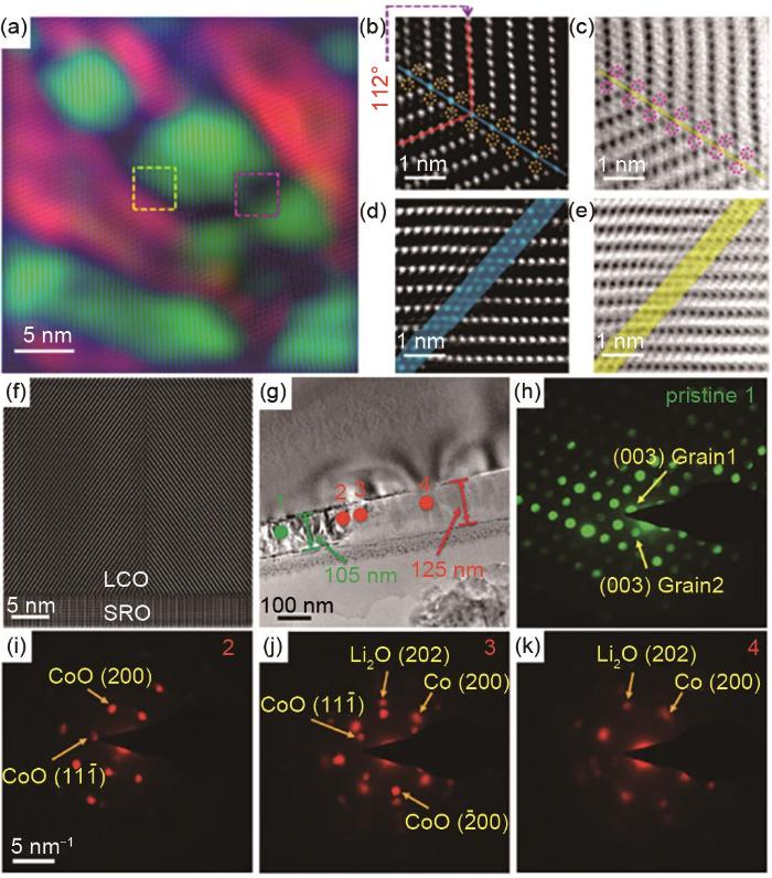
... [37];(f) 原始LCO薄膜样品的HAADF-STEM图;(g) 嵌锂后的低倍TEM图;(h)~(k) 在(g)中标记不同点的NBD图[38]<strong>(a) HAADF image of delithiated LiCoO<sub>2</sub> cathode after coloring with GPA method, (b) and (c) are zoomed-in views of yellow, dashed-box area, (d) and (e) are zoomed-in images of pink, dashed-box area</strong><sup>[<xref ref-type="bibr" rid="R37">37</xref>]</sup><strong>, (f) A HAADF-STEM image of a pristine thin film LCO piece, (g) A TEM image after lithiation acquired at a low magnification, (h)</strong>~<strong>(k) NBD patterns for different spots labeled in (g)</strong><sup>[<xref ref-type="bibr" rid="R38">38</xref>]</sup>Fig. 1
... [37], (f) A HAADF-STEM image of a pristine thin film LCO piece, (g) A TEM image after lithiation acquired at a low magnification, (h)~(k) NBD patterns for different spots labeled in (g)[38]Fig. 1
<strong>(a) HAADF image of delithiated LiCoO<sub>2</sub> cathode after coloring with GPA method, (b) and (c) are zoomed-in views of yellow, dashed-box area, (d) and (e) are zoomed-in images of pink, dashed-box area</strong><sup>[<xref ref-type="bibr" rid="R37">37</xref>]</sup><strong>, (f) A HAADF-STEM image of a pristine thin film LCO piece, (g) A TEM image after lithiation acquired at a low magnification, (h)</strong>~<strong>(k) NBD patterns for different spots labeled in (g)</strong><sup>[<xref ref-type="bibr" rid="R38">38</xref>]</sup>Fig. 1
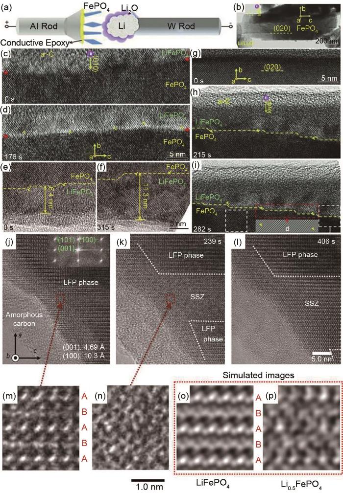
... [42];(j)~(l)为LFP纳米线在嵌锂过程中的微观结构演变图;(m)、(n) 在(j)、(k)中红色虚线矩形图像的放大图;(o)和(p)为LFP和Li0.5FePO4的TEM模拟图[43]<strong>(a) Schematic diagram of <i>in situ</i> TEM setup with nanobattery, (b) A TEM image of FePO<sub>4</sub> particle after its contact with Li/Li<sub>2</sub>O solid electrolyte, (c)</strong>~<strong>(i) Time-lapse TEM images of FePO<sub>4</sub> particle during lithiation</strong><sup>[<xref ref-type="bibr" rid="R42">42</xref>]</sup><strong>, (j)</strong>~<strong>(l) Microstructural evolution of a LFP nanowire during lithiation, (m)</strong>、<strong>(n) Magnified images of dotted red rectangle region in (j)</strong>、<strong>(k), respectively. Simulated TEM images of (o) LiFePO<sub>4</sub> and (p) Li<sub>0.5</sub>FePO<sub>4</sub></strong><sup>[<xref ref-type="bibr" rid="R43">43</xref>]</sup>Fig. 2
... [42], (j)~(l) Microstructural evolution of a LFP nanowire during lithiation, (m)、(n) Magnified images of dotted red rectangle region in (j)、(k), respectively. Simulated TEM images of (o) LiFePO4 and (p) Li0.5FePO4[43]Fig. 2
<strong>(a) Schematic diagram of <i>in situ</i> TEM setup with nanobattery, (b) A TEM image of FePO<sub>4</sub> particle after its contact with Li/Li<sub>2</sub>O solid electrolyte, (c)</strong>~<strong>(i) Time-lapse TEM images of FePO<sub>4</sub> particle during lithiation</strong><sup>[<xref ref-type="bibr" rid="R42">42</xref>]</sup><strong>, (j)</strong>~<strong>(l) Microstructural evolution of a LFP nanowire during lithiation, (m)</strong>、<strong>(n) Magnified images of dotted red rectangle region in (j)</strong>、<strong>(k), respectively. Simulated TEM images of (o) LiFePO<sub>4</sub> and (p) Li<sub>0.5</sub>FePO<sub>4</sub></strong><sup>[<xref ref-type="bibr" rid="R43">43</xref>]</sup>Fig. 2
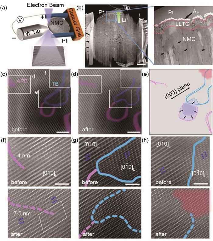
... [44]<strong>(a) Schematic of <i>in situ</i> TEM experimental setup; (b) low magnification HAADF-STEM images; (c),(d)HR-HAADF-STEM images of NMC before and after delithiation, respectively; (e) Phases extension and defects migration; (f)</strong>~<strong>(h) Magnified images of (c),(d)</strong><sup>[<xref ref-type="bibr" rid="R44">44</xref>]</sup>Fig. 3
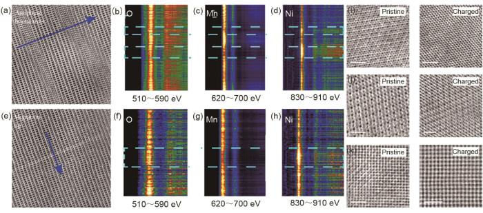
... [45]<strong>(a) HAADF-STEM image of antiphase boundaries, (b)</strong>~<strong>(d) EELS spectrum of O, Mn, and Ni from (a), respectively, (e) HAADF-STEM image of migration-front area, (f)</strong>~<strong>(h) EELS spectrum of O, Mn, and Ni from (e), (i)</strong>~<strong>(k)ABF-STEM images of pristine and charged LNMO along [111], [110] and [100] direction, respectively</strong><sup>[<xref ref-type="bibr" rid="R45">45</xref>]</sup>Fig. 42 锂离子电池负极材料的原位TEM研究
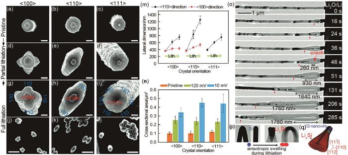
... [55];(o), (p) Si纳米线在锂化过程中微结构演变的TEM图;(q) 逐步锂化的Si纳米线的三维模拟图[56]<strong>(a)</strong>~<strong>(l) Top-view SEM images of Si nanopillars along [100], [110] and [111] directions at different lithiation states; (m), (n) Statistical data of changes in cross-sectional dimensions and cross-sectional area along these three orientations</strong><sup>[<xref ref-type="bibr" rid="R55">55</xref>]</sup><strong><sup>;</sup> (o), (p) Microstructural evolution of Si nanowire during lithiation; (q) 3D simulation plot of a progressively lithiated Si nanowire</strong><sup>[<xref ref-type="bibr" rid="R56">56</xref>]</sup>Fig. 5
... [55]; (o), (p) Microstructural evolution of Si nanowire during lithiation; (q) 3D simulation plot of a progressively lithiated Si nanowire[56]Fig. 5
... [56]<strong>(a)</strong>~<strong>(l) Top-view SEM images of Si nanopillars along [100], [110] and [111] directions at different lithiation states; (m), (n) Statistical data of changes in cross-sectional dimensions and cross-sectional area along these three orientations</strong><sup>[<xref ref-type="bibr" rid="R55">55</xref>]</sup><strong><sup>;</sup> (o), (p) Microstructural evolution of Si nanowire during lithiation; (q) 3D simulation plot of a progressively lithiated Si nanowire</strong><sup>[<xref ref-type="bibr" rid="R56">56</xref>]</sup>Fig. 5
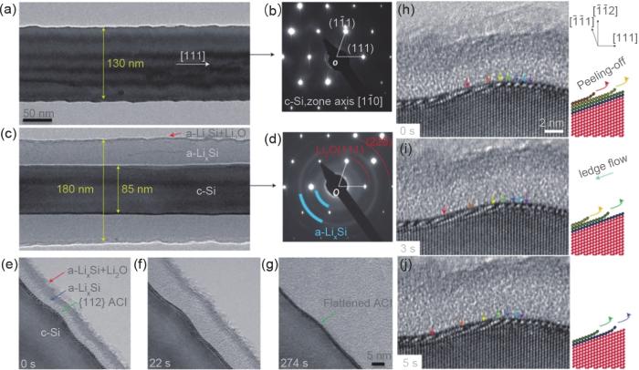
... [57]<strong>(a)</strong>~<strong>(d) TEM images and corresponding electron diffraction patterns of a pristine silicon nanowire and a partially lithiated silicon nanowire; (e)</strong>~<strong>(g) Time-lapse images showing migration of ACI during lithiation; (h)</strong>~<strong>(j) TEM images and diagrams of lithiation process by lateral ledge flow in ACI and simultaneous peeling-off</strong><sup>[<xref ref-type="bibr" rid="R57">57</xref>]</sup>Fig. 6
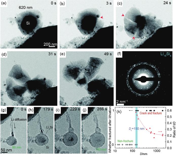
... [58]<strong>(a)</strong>~<strong>(e) TEM images showing fast fracture of 620 nm Si nanoparticle during lithiation process. (f) Electron diffraction pattern of broken Si nanoparticle after lithiation, indicating formation of Li<sub>15</sub>Si<sub>4</sub>. (g)</strong>~<strong>(j) TEM images of a 80 nm Si nanoparticle without cracking during lithiation. (k) Statistics showing critical size of Si nanoparticles without cracking during lithiation process</strong><sup>[<xref ref-type="bibr" rid="R58">58</xref>]</sup>Fig. 7
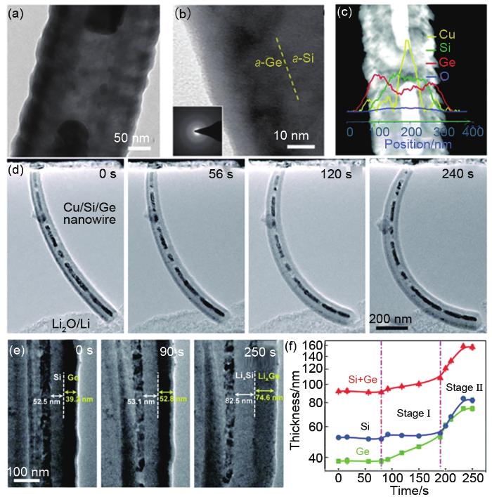
... [60](a) TEM image of a Cu/Si/Ge nanowire; (b) HRTEM image of Si/Ge bilayer and corresponding SAED pattern; (c) STEM image of Cu/Si/Ge nanowire and corresponding EDX spectra; (d), (e) Time-lapse TEM images of Cu/Si/Ge nanowire during lithiation; (f) Evolution of layer thickness in Si/Ge bilayer shell during lithiation<sup>[<xref ref-type="bibr" rid="R60">60</xref>]</sup>Fig. 8
... [65]<strong>(a)</strong>~<strong>(f) Time-lapse TEM images of AMPSi@C electrode during lithiation. <i>In situ</i> TEM images of AMPSi@C (g) after 1st cycle, (h) after 2nd cycle, (i) after 3rd cycle and (j) after 4th cycle</strong><sup>[<xref ref-type="bibr" rid="R65">65</xref>]</sup>Fig. 9
... [73]<strong>(a)</strong>~<strong>(c) Time-lapse TEM images of a yolk@shell Fe<sub>2</sub>O<sub>3</sub>@C electrode during first lithiation process; TEM images acquired after first lithiation (d), and first delithiation (h); SAED patterns before lithiation (e), after first lithiation (f) and after first delithiation (g); TEM images after second lithiation (i), after second delithiation (j), after third lithiation (k) and after third delithiation (l)</strong><sup>[<xref ref-type="bibr" rid="R73">73</xref>]</sup>Fig. 10
... [74]<strong>(a)</strong>~<strong>(c) Time-resolved TEM images of a FeP@CNs electrode during lithiation. <i>In situ</i> TEM images and corresponding SAED patterns of pristine FeP@CNs electrode (d)</strong>~<strong>(g), after first lithiation [(e), (h)] and first delithiation [(f), (i)]</strong><sup>[<xref ref-type="bibr" rid="R74">74</xref>]</sup>Fig. 11


